Visit UCR Return to Campus website - Take the COVID Screening Check survey
Under an optical microscope, carbon nanotubes, for example, look grayish. The inability to distinguish fine details and differences between individual pieces of nanomaterials makes it hard for scientists to study their unique properties and discover ways to perfect them for industrial use.
In a new report in Nature Communications, researchers from UC Riverside describe a revolutionary imaging technology that compresses lamp light into a nanometer-sized spot. It holds that light at the end of a silver nanowire like a Hogwarts student practicing the “Lumos” spell, and uses it to reveal previously invisible details, including colors.
The advance, improving color-imaging resolution to an unprecedented 6 nanometer level, will help scientists see nanomaterials in enough detail to make them more useful in electronics and other applications.
Ming Liu and Ruoxue Yan, associate professors in UC Riverside’s Marlan and Rosemary Bourns College of Engineering, developed this unique tool with a superfocusing technique developed by the team. The technique has been used in previous work to observe the vibration of molecular bonds at 1-nanometer spatial resolution without the need of any focusing lens.
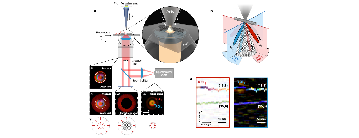
In the new report, Liu and Yan modified the tool to measure signals spanning the whole visible wavelength range, which can be used to render the color and depict the electronic band structures of the object instead of only molecule vibrations. The tool squeezes the light from a tungsten lamp into a silver nanowire with near-zero scattering or reflection, where light is carried by the oscillation wave of free electrons at the silver surface.
The condensed light leaves the silver nanowire tip, which has a radius of just 5 nanometers, in a conical path, like the light beam from a flashlight. When the tip passes over an object, its influence on the beam shape and color is detected and recorded.
The light is then focused into a spectrometer, where it forms a tiny ring shape. By scanning the probe over an area and recording two spectra for each pixel, the researchers can formulate the absorption and scattering images with colors. The originally grayish carbon nanotubes receive their first color photograph, and an individual carbon nanotube now has the chance to exhibit its unique color.
The new technology can be an important tool to help the semiconductor industry make uniform nanomaterials with consistent properties for use in electronic devices. The new full-color nano-imaging technique could also be used to improve understanding of catalysis, quantum optics, and nanoelectronics.
Liu, Yan, and Ma were joined in the research by Xuezhi Ma, who worked on the project as part of his doctoral research at UCR Riverside. Researchers also included UCR students Qiushi Liu, Ning Yu, Da Xu, Sanggon Kim; Zebin Liu and Kaili Jiang at Tsinghua University, and UCR professor Bryan Wong. The paper, titled “6 nm super-resolution optical transmission and scattering spectroscopic imaging of carbon nanotubes using a nanometer-scale white light source,” is available here.
Research highlight from the UCR News: https://news.ucr.edu/articles/2021/11/23/magic-wand-reveals-colorful-nano-world